Radiation exposure of plane x-ray of the sinuses is 0. 3msv, while radiation exposure of ct scan of the sinuses is 0. 7msv, so ct scan is also considered a low radiation exposure compared to abdominal ct scans which can reach 8. 00msv, this is the difference in the radiation exposure that most doctors are claiming to safe the patient by not getting them exposed to it, but they forget that they. Continued. computed tomography : a ct scanner uses x-rays and a computer to create detailed images of the sinuses. ct scanning can help diagnose chronic sinusitis. magnetic resonance imaging. Don't delay your care at mayo clinic featured conditions see our paling aman precautions in response to covid-19. request an appointment. a chest x-ray helps detect problems with your heart and lungs. the chest x-ray on the left is normal. the i. Fever, headache, postnasal discharge of thick sputum, nasal congestion and an abnormal sense of smell. acute sinusitis is a clinical diagnosis characterized by symptom duration of less than 4 weeks 11.
Noncontrast coronal computed tomographic (ct) images can define the nasal anatomy much more precisely. mucosal thickening, polyps, and other sinus abnormalities can be seen in 40 percent of. X-rays are a common diagnostic tool for identifying broken bones, which may first come to mind, but it’s also useful in evaluating the possibility of sinusitis. sinusitis is inflammation or infection in the sinus facial cavity, the area surrounding the nose, eyes, and cheeks inside the skull. Learn more about x-rays, a quick and painless, non-invasive diagnostic tool used to x images ray sinusitis detect and stage cancerous tumors. the information on this laman was reviewed and approved by maurie markman, md, president, medicine & science at ctca. this.
What Is A Sinus Xray Two Views

This laman explains the different types of medical imaging procedures that are used on children. it includes reasons why unnecessary radiation exposure during medical procedures should be minimized in pediatric patients. the. gov means it’s. Chronic sinusitis is defined clinically as a sinonasal infection lasting more than 12 weeks. patients may present with symptoms of sinusitis such as nasal obstruction, nasal discharge, facial pain, headache, halitosis, anosmia, etc. it is worth noting is no definite correlation between symptoms and imaging findings of chronic sinusitis and that endoscopic chronic sinusitis may have no imaging correlation as the mucosa is best appreciated on the former 11.
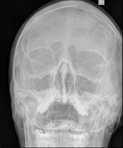
Johns hopkins medical imaging provides x-ray procedures at convenient locations in green spring station, white marsh, columbia and bethesda. due to interest in the covid-19 vaccines, we are experiencing an extremely high call volume. please. Sinus x-rays may appear normal or show infection through mucosal thickening, fluid levels, or total opacity(9). an opaque sinus may be caused by the thickening of the bony walls, small asymmetric antra, or improper centering and rotation of the head. this type of sinus can pose difficulties in interpreting radiological appearance.
Sinus Xray Photos Free Royaltyfree Stock Photos From Dreamstime

Sinus Xray Photos Free Royaltyfree Stock Photos From
Li z, wang x, jiang h, qu x, wang c, chen x, et al. chronic invasive fungal rhinosinusitis vs sinonasal squamous cell carcinoma: the differentiating value of mri. eur radiol. 2020 aug. 30 (8):4466-4474. jeon y, lee k, sunwoo l, et al. deep learning for x images ray sinusitis diagnosis of paranasal sinusitis using multi-view radiographs. These x-ray pictures explore the human body, diseases, conditions and implants. see x-ray pictures to see the amazing images of the human body. advertisement by: discovery fit and health writers two surgeons examine an x-ray of a broken leg. A sinus x-ray involves the use of radiation to create images of your body. while it uses relatively low amounts of radiation, there is still a risk every time your body is exposed to radiation. More sinusitis x ray images images.
Computed tomography (ct) scanning is the examination of choice in sinusitis, particularly in cases of chronic sinus disease, providing excellent detail of sinus anatomy. however, ct is usually not useful in acute sinusitis, as penaksiran in acute cases is primarily based on clinical findings. good anatomic definition is desirable before surgical intervention. [14, 15, 16, 17, 18, 19] coronal ct imaging is the preferred initial procedure. bone-window views provide excellent resolution and good definition of the complete ostiomeatal complex and other anatomic details that play a role in sinusitis. in addition, the coronal view is best correlated with findings from sinus surgery, with anatomy and pathology visualized in a plane almost identical to that seen by the endoscopist. ct provides an excellent anatomic display of soft-tissue attenuation. this depiction includes fluid levels and polypoid masses within the normally air-filled cavities of the sinuses, nasal cavity, and postnasal spa X-ray film of the face frontal, nose-chin and lateral projection. volume formation of the right maxillary sinus. marker. negativ. a images of the x-ray face. A characteristic feature on ct sinuses is sclerotic thickened bone (hyperostosis) involving the sinus wall from a prolonged mucoperiosteal reaction. intrasinus calcificationmay be present. the presence of opacification is not a good discriminator from an acute sinus infection. there are five main patterns x images ray sinusitis of chronic inflammatory disease that classify the disease into distinct anatomical/pathological groups and are dependent on the drainage pathways affected. this classification helps the surgeon to select the type of surgery needed 12: 1. ostiomeatal complex pattern: maxillary sinus, anterior ethmoid air cells, and frontal sinuses are affected due to obstruction of the ostiomeatal complex dua. infundibular pattern: isolated obstruction to the ethmoid infundibulum and/or maxillary sinus ostium tiga. sphenoethmoidal recess pattern: inflammatory changes in the sphenoethmoidal recess obstruct the sphenoid sinus in isolation or in conjunction with the posterior ethmoidal air cells 4. sinonasa
See full list on radiopaedia. org. Conservative medical treatment until the inflammation subsides and treatment of the cause, e. g. dental caries. if it becomes chronic sinusitis, functional endoscopic sinus surgerymay be considered. 1. erosion through bone 1. 1. subperiosteal abscess 1. 1. 1. frontal sinus superficially (pott puffy tumor) 1. 1. dua. frontal or ethmoidal sinuses into the orbit (subperiosteal abscess of the orbit) dua. dural venous sinus thrombosis tiga. intracranial extension tiga. 1. meningitis 3. 2. subdural empyema tiga. tiga. cerebral abscess. Sinus x-rays may appear normal or show infection through mucosal thickening, fluid levels, or total opacity (9). an opaque sinus may be caused by the thickening of the bony walls, small asymmetric antra, or improper centering and rotation of the head. this type of sinus can pose difficulties in interpreting radiological appearance. Computed tomography (ct) scanning is the examination of choice in sinusitis, particularly in cases of chronic sinus disease, providing excellent lebih jelasnya of sinus anatomy. x images ray sinusitis the ostiomeatal units are brilliantly shown on ct scans, which provide greater definition of the pathology than do other images, especially within the sphenoid and ethmoid sinuses.
To evaluate the pattern of sinusitis, one must understand the drainage of various sinuses. the anatomy of drainage revolves around the ostiomeatal unit, which is not a single morphologic structure but a combination of the following structures: 1. middle turbinate 2. ethmoid bulla 3. uncinate process 4. maxillary infundibulum lima. hiatus semilunaris (ie, space beneath the middle turbinate) 6. maxillary os the hiatus semilunaris is a space between the uncinate process (anteroinferiorly) and the ethmoid bulla (posterosuperiorly). the anterior group of ethmoid air cells drains into the anterior aspect of the hiatus semilunaris through the frontonasal duct. the middle group drains into the hiatus semilunaris on or above the ethmoidal bulla. the frontal sinus drains through the frontonasal duct or through the anterior ethmoidal cells into the hiatus semilunaris. the maxillary infundibulum drains into the posterior part of the hiatus semilunaris. the frontal, maxillary, anterior, and middle See full list on emedicine. medscape. com.
Nov 15, 2002 · noncontrast coronal computed tomographic (ct) images can define the nasal anatomy much more precisely. mucosal thickening, polyps, and other sinus abnormalities can be seen in 40 percent of. The american college of radiology (acr) regards noncontrast ct scanning as the examination of choice in recurrent or chronic sinus disease. all imaging findings are interpreted in conjunction with clinical and endoscopic findings. mri is a complementary study when aggressive disease is being evaluated with ophthalmic/intracranial complications, especially in fungal disease in immunocompromised patients and in characterization of a sinus mass. [28] the acr recommendations for sinusitis in children include the following[29] : 1. imaging studies are not recommended for uncomplicated acute sinusitis: 2. ct of the paranasal sinuses without iv contrast is recommended for persistent sinusitis (worsening course or severe presentation, or not responding to treatment), recurrent sinusitis, or chronic sinusitis, or to define paranasal sinus anatomy before functional endoscopic sinus surgery. 3. ct or mri of the head and paranasal sinuses with iv contrast is recommended for sinusitis with clinic
Posting Komentar untuk "X Images Ray Sinusitis"